


Digitalization has made medical imaging more accessible and faster. AI is increasingly improving the process, making medical imaging an essential tool for healthcare professionals worldwide.
In this article, we will explore the world of medical imaging along with Artificial Intelligence (AI) in medical image processing. We will also learn how AI is revolutionizing the field of medicine. We will delve into the methods AI is being utilized to enhance medical imaging techniques, improve diagnosis, and ultimately save lives. Dive in to learn more.
To start from the very beginning, let us discuss the description of an image in terms of computing.
A computer sees the image as a two-dimensional function F(x,y), where x and y are spatial coordinates, and the amplitude of F at any pair of coordinates (x,y) represents the intensity of that image at a specific point. A digital image consists of a number of elements, each with a particular value at a particular location, known as pixels. A pixel can be characterized by its shade, opacity, or color. The following scales are usually used to represent the pixels:
This type of image has only two unique values for pixel intensity, either 0 or 1, which represent black and white, respectively. It is commonly used in biomedical image segmentation.
Grayscale images have 256 unique shades of gray, with 0 representing black and 255 representing white. All the other values in between are different shades of gray. Converting an RGB image to greyscale results in the same shape of histogram (a graphical representation of data using bars of different heights).
Most images that we see in daily life are RGB or colored images. To machines, these are 16-bit matrices with 65,536 different colors possible for each pixel. RGB stands for red, green, and blue channels in an image. Each channel has values ranging from 0 to 255, and three equal-sized matrices are stacked on top of each other to form a pixel in the image. The different combinations of values for each channel result in all the colors in nature.
This type of image is a colored RGB image with an extra channel called “alpha,” which represents the opacity of the image. Opacity ranges from 0% to 100% and mimics the “see-through” property of objects in real life.
Images are presented as a two-dimensional array arranged in rows and columns or, in other words, as a matrix, with each element of the matrix representing a pixel.
Digital image processing involves converting analog visuals (for example, from X-rays or CT in medical imaging) into digital format for further use in diagnosis.
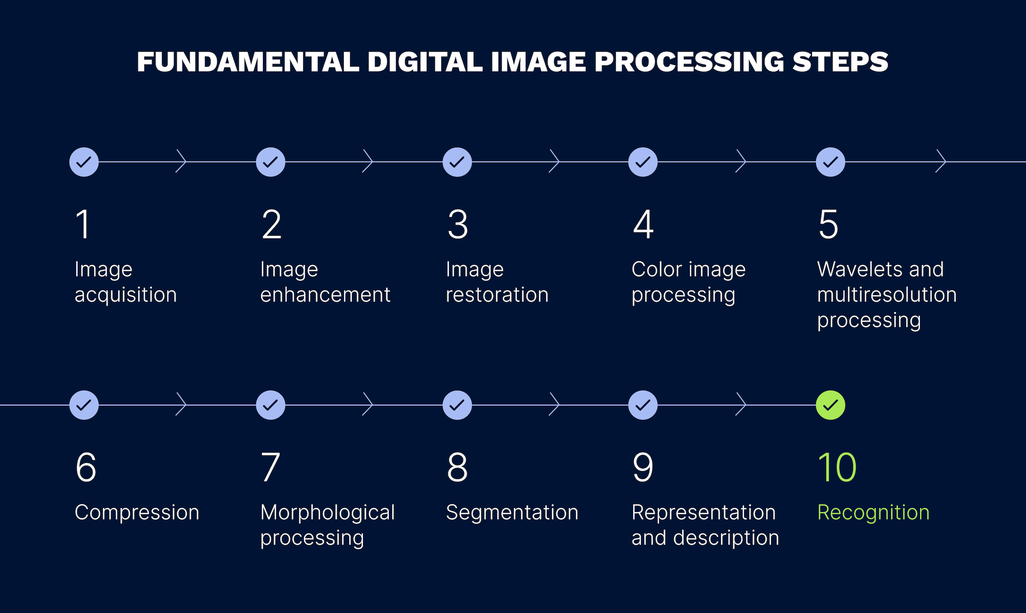
Image processing comprises various steps that facilitate the handling of digital images. Here are the fundamental steps of digital image processing:
Image acquisition. This step involves pre-processing, where an image is retrieved from a hardware-based source.
Image enhancement. The process of enhancing an image involves bringing out and highlighting certain features of interest by adjusting brightness, contrast, or other image characteristics.
Image restoration. Image restoration improves the appearance of an image based on specific mathematical or probabilistic models.
Color image processing. Color image processing includes various image processing techniques for modeling color in digital images.
Wavelets and multi-resolution processing. This step subdivides images into wavelets or smaller regions for data compression and pyramidal representation.
Compression. Compression is used to reduce the storage required to save images or the bandwidth required to transmit them, especially for use on the internet.
Morphological processing. Morphological processing uses specific algorithms to morph images based on their shapes.
Segmentation. Segmentation involves partitioning an image into its constituent parts or objects, making it easier to analyze and manipulate.
Representation and description. After segmentation, image regions are represented and described in a form suitable for further computer processing.
Recognition. Recognition involves assigning a label to an object based on its description.
Let us now turn to medical imaging and briefly describe the field in order to provide a smooth transition into the context that follows.
Here are the types of commonly used medical imaging systems:
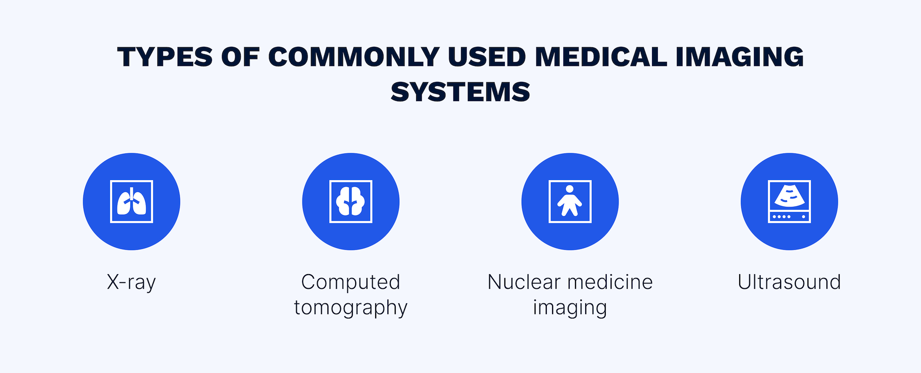
Since the German scientist W.C. Röntgen discovered X-rays, they have been used to image body parts for diagnostic purposes.
This is how it works: the electron beam is generated by a thermal emission process and accelerates through a potential difference of 50-150 kV. To produce X-rays, the electrons strike the anode whereby only 1% of this energy is converted into X-rays; the rest is converted into heat. X-ray machines produce 2D plans of the examined parts of the body. Therefore, to evaluate a case, 2 different views are often required.
Unlike conventional radiography, CT produces images in multiple dimensions. The CT scanner generates multiple slices of the body tissues in different directions. In the CT scanner, the patient is placed in the aperture and scanned in all directions by a rotating X-ray tube. It can produce a detailed cross-sectional image of the bone, blood, vessel, and soft tissues. Medical professionals commonly use CT scans to evaluate complex regions of the body, such as spine or pelvis.
This imaging technique uses radioactive isotopes to produce images of the function of different structures, such as the heart, kidneys, and liver. The radioactive isotopes are labeled with pharmaceutical materials so that they can be directed to the specific organs. The detectors receive the photons emitted by the patient and convert them into signals. These signals are then transformed into interpretable digital images. There are several types of nuclear medicine scanning modalities, such as planar, tomographic, and positron emission. Planar emission produces 2D images. Both tomographic and positron emission produce 3D images.
Ultrasound is a technique that uses high frequency sound waves to create images of the internal structure of the body from the echoes that are returned to it. Ultrasound is similar to the locating technique that is used by some animals, such as bats and whales, in the natural world. Ultrasonic waves are transmitted into the body in radio frequency pulses through a transducer while these waves pass through the body tissue. Some of these waves are absorbed and some are reflected back. The reflected waves are received by the transducer and converted into electrical signals. These electrical signals are then converted into digital signals and run through the computer system. The computer system uses arithmetic and logic calculations to create a 2D image of the scanned structures. In the ultrasound system, thousands of pulses are sent every millisecond. Many imaging techniques are used to enhance the ultrasound images.
Despite the advances of medical imaging, its interpretation and analysis remain a significant challenge. The causes of many related problems recognized by medical professionals include:
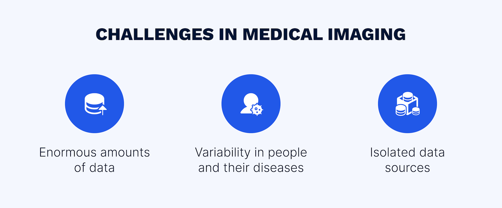
The examples of challenges in medical image processing
People and their diseases differ despite more than 90 percent of humans share the same genetic material. Even though numerous cases are classified, the classification is not specific enough due to variabilities within the same disease, which can be seen on a molecular level. In addition to molecular diversity, there is phenotypic diversity which leads to variable lesions on images. Additionally, there are clinical differences between the patients in terms of how they respond to treatment.
Growth in electronic patient data is a setup for errors. Clinicians require computer assistance to interpret an increasing number of images together with non-image data. Due to digitization and storing data in electronic format, there are many opportunities to use this data for building and training models to discover the best treatments for patients.
Today’s clinicians are faced with excessive amounts of data and increasingly complex datasets that include not only radiological information, but also genomic data, pathology reports and blood analyses. There has also been a dramatic increase in therapeutic options, particularly in oncology. Therefore, integrating various types of data (for example, images and clinical notes) is of urgent need. Through the integration of multiple data sources (including 2D and 3D medical images, ECGs, EEGs, EMGs, and EHRs, vital signs such as body temperature, pulse rate, respiratory rate, blood pressure, demographic information, medical history, and lab test results), healthcare providers can have a comprehensive understanding of a patient’s health and the underlying cause s of their symptoms. In addition, reduced risk of misdiagnosis and improved diagnostic accuracy can be achieved.
The issues described above lead to the following consequences:
And that’s where AI comes in. With its remarkable ability to learn, recognize patterns, and analyze complex data sets, AI is drastically changing medical imaging by providing solutions to some of the most essential issues.
AI is essentially the capability of computers to learn, and that learning can be supervised, unsupervised, or semi-supervised.
Supervised learning includes but is not limited to algorithmic (involves the use of specific sequences of steps or algorithms like decision trees, logistic regression, or neural networks) or topological (involves the use of topological data analysis (TDA) to identify patterns and relationships in high-dimensional data) methods. In supervised learning, algorithms learn from labeled data. In case of medical imaging, medical experts have to label a vast number of images to highlight the area of interest.
For semi-supervised learning, medical professionals only need feedback from software confirming that it works.
The main tasks of unsupervised learning are clusterization, which is essentially grouping things together, and search. For unsupervised learning, raw data that is represented by unlabeled images can be used.
AI in medical imaging refers to the edge of artificial intelligence: machine learning, deep learning, and artificial neural networks.
To better understand how these technologies work, we first need to look at traditional programming. In traditional programming, medical software developers write code to instruct a machine to perform certain tasks. The machine gets better at these tasks by following the same set of instructions repeatedly over time. Machine learning in healthcare and other industries, on the other hand, uses algorithms that allow machines to learn from data and to improve their performance over time. Rather than being given specific instructions on how to perform a task, the machine is fed large amounts of data. It then uses that data to identify patterns and relationships that can be used to make predictions or decisions about the data. Deep learning is a subfield of machine learning that uses artificial neural networks (ANNs). ANNs are patterned after the structure and function of the human brain. They are made up of layers of interconnected nodes that process information and pass it on to the next layer of nodes. Each node in an ANN performs a simple calculation, but the combination of all the nodes in a layer results in a complex output. Deep learning in medical image analysis uses multi-layer ANNs to learn more abstract features from data.
The basic areas where AI is currently playing an essential role are described below.
AI is transforming the field of medical imaging in countless ways. Here are some of the most promising applications of AI in biomedical imaging:
Reading and interpreting images are the areas in which AI is extensively used in radiology. Radiologists review numerous images each day in a high-pressure environment. AI-based clinical decision support can aid in spotting suspicious elements without having to report false positives.
The digital images that radiologists review are produced in a standard DICOM (digital imaging and communication in medicine) format, accessed through the picture archiving communication system (PACS). The standardized nature of these images and the PACS make it simpler for third parties to use the data for algorithm development. They can then make those algorithms available to the widest customer base possible, as they can be used regardless of the system capturing the image. Triage tools are popular among AI algorithm developers as they can help radiologists prioritize which images to focus on first, particularly in time-sensitive cases such as strokes. Several AI applications have received FDA clearance for support tools that triage potentially life-threatening diagnosis, including stroke, intracranial hemorrhage, pulmonary embolism, and spine fractures.
Software solution for analyzing, transmitting, and viewing medical imaging data in the DICOM format.
Post-scanning image reconstruction is a key area that benefits from AI. Deep-learning algorithms that increase signal-to-noise ratios are typically used to improve image reconstruction. This leads to shorter scan times, increased diagnostic confidence, and accelerated workflows, resulting in images that are 30% faster, have higher quality, and more resolution. With this medical image processing software, you can significantly increase the number of patients scanned per day from 20-25 to 30-35.
Another area where AI image processing is used in the medical field is segmentation, which involves the AI system analyzing the scan and measuring the volume of an organ or abnormality. When performed manually, medical image segmentation is a time-consuming task that divides the image into parts that represent normal tissue and those that are abnormal. AI significantly reduces the time required for this activity.
The convolutional neural network U-Net is a specialized tool for the automated segmentation of medical images, including those of the brain and liver. Such segmentation improves image analysis and provides radiologists with the confidence to make a diagnosis.
Image registration involves aligning multiple images of the same patient or organ to enable consistent comparison and analysis. AI algorithms are increasingly being used to automate the process of image registration, improving the accuracy and reliability of this critical task.
Another important application of AI in medical image processing is image enhancement. Medical images must often be captured under less-than-optimal conditions, with limited resolution, contrast, or clarity. AI algorithms can be trained to recognize and enhance key features within these images, such as blood vessels or tumors, making them easier for physicians to detect and analyze.
Yet another area in which AI is transforming medical image processing is real-time image analysis. With AI algorithms running in the background, imaging equipment can now produce incredibly detailed, real-time images during procedures such as surgery. This allows doctors and other medical professionals to make faster, more informed decisions, leading to improved patient outcomes and more efficient use of resources.
The advances in AI in medical image processing are set to revolutionize the healthcare industry, providing doctors with more accurate and effective tools for diagnosing and treating patients. As the technology continues to mature and evolve, we can expect even more exciting applications of AI in medical imaging.
AI has proved to have many benefits for medical imaging, yet it is still often seen as a technology of the future rather than a real-time solution. However, by implementing the appropriate AI-backed solution, many doubts can be eliminated. This is where EffectiveSoft comes in. With our extensive experience in AI, ML, and computer vision end-to-end software development for the healthcare domain, and our team of top-notch engineers, we are well-equipped to streamline your healthcare operations.
Don’t hesitate to contact us. We are happy to help with your healthcare project by leveraging the power of AI.
Blog


Our team would love to hear from you.
Fill out the form, and we’ve got you covered.
What happens next?
San Diego, California
4445 Eastgate Mall, Suite 200
92121, 1-800-288-9659
San Francisco, California
50 California St #1500
94111, 1-800-288-9659
Pittsburgh, Pennsylvania
One Oxford Centre, 500 Grant St Suite 2900
15219, 1-800-288-9659
Durham, North Carolina
RTP Meridian, 2530 Meridian Pkwy Suite 300
27713, 1-800-288-9659
San Jose, Costa Rica
C. 118B, Trejos Montealegre
10203, 1-800-288-9659

